Positron Emission Tomography (PET/CT)
Positron emission tomography is a method of imaging used to determine the level of functioning in tissues and organs. A radioactive substance is used to determine how tissues and organs function. A wide variety of radioactive materials can be used for this purpose, and they can be administered through the mouth or a vein. Among radioactive substances, F-18 FDG - a glucose molecule labeled with radioactive substance- is the most common one. F18-FDG is administered into a vein, and the uptake of this substance by tissues is checked. Since malignant cells show high affinity, the activity level in cancer cells is demonstrated.
Computed tomography is a radiological modality that allows the cross-sectional imaging of the body.
When these two screening methods are combined, PET / CT takes the cross-sectional views of the target region of the body, and the activity in the tissues and organs is traced. For this reason, the radioactive substance used in PET / CT is called a "tracer." Thus, it is possible to obtain detailed information about the location and the activity level of the diseased area by combining the clear cross-sectional images with the images that show the activity uptake (the color tone or brightness level according to the extent of uptake of radioactive substance).
PET scan, using the activity uptake feature of tissues, is helpful in the diagnosis, treatment, and follow-up of cancers, heart diseases (coronary artery disease, heart attack), and brain diseases (brain tumor, epileptic seizures). The disorders that PET / CT can diagnose are listed below:
1. Cancers
- Brain
- Head and neck
- Esophagus
- Lung
- Colon and rectum
- Pancreas
- Prostate
- Thyroid
- Cervix
- Lymphoma
- Melanoma
2. Investigating viable tissue after coronary artery disease, coronary artery bypass grafting and heart attack
3. Brain tumors
4. Differential diagnosis of Alzheimr Disease and dementia
5. Epileptic seizuresIf PET/CT is used for cancer screening, your cancer will be screened at intervals that your doctor will determine and instruct.
In other words, after the diagnosis of cancer is established and the treatment is started, the response of cancer to the treatment is assessed. In the light of this response, the dose can be modified, additional medications can be added, active medications can be stopped, and another protocol can be started, or it can be decided to maintain the treatment.
As PET/CT images the tissue activity, it guides, by reflecting the amount of viable tissue in the heart, to the determination of the most appropriate treatment for the patient in cases of heart attack, coronary artery disease, etc.
For central nervous system disorders, it is used for the differential diagnosis of Alzheimer's disease, and it may play an essential role in clarifying the origin of epileptic seizures.
If your doctor thinks a PET/CT is necessary or valuable, an appointment is scheduled for this screening. After the appointment is scheduled, you are usually given a brochure, where you can see what you need to do before the appointment date. Due to exposure to radiation (X-ray) during PET / CT scans, if you are pregnant or breastfeeding, you should inform your doctor and healthcare personnel.
You will be asked not to eat or drink (except water) for at least 6 to 8 hours before PET/CT. It will also be questioned whether or not you have experienced an allergic reaction to contrast agents in the past.
Since 18F-FDG is a sugar molecule that is most commonly used as a tracer for PET / CT imaging, you will be assessed explicitly for diabetes mellitus. Moreover, after your medicine is administered, your blood sugar will be measured before the scan is initiated.
When a PET / CT scan is scheduled, you need to notify your doctor and healthcare professionals about all your prescription and over-the-counter medicines, herbal products, and vitamin and mineral supplements that you use.
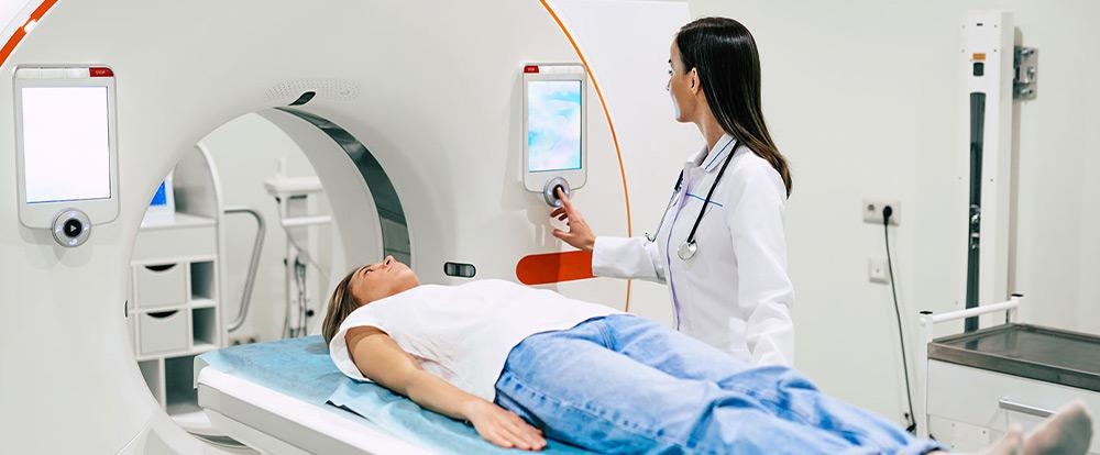
After the radioactive substance to be used for PET/CT imaging is administered through the mouth or a vein (intravenous), you will be asked to relax in a room for about 1 hour to ensure that the radioactive substance is homogeneously distributed throughout the body.
After this time elapses, you will be transferred to the room, where images will be obtained. If you have had an MRI or CT scan in the past, this environment will be familiar to you. (claustrophobia), informing your doctor before the PET/CT scan may be helpful for the smooth completion of the scan. The imaging machine will produce bizarre sounds while your body images are obtained. Once the scanning is completed, you can leave the hospital without any exceptional advice if you are not already hospitalized. Drinking plenty of fluids after the scan will help to accelerate the excretion of the radioactive material. Radiologists will review the images and issue a report according to the indication of the scanning (diagnosis and follow-up of cancer, response to treatment in cancer, heart diseases, and central nervous system disorders). When your report is available to the doctor who requested the PET/CT, your diagnosis, treatment, and follow-up period will be planned, and you will be informed.